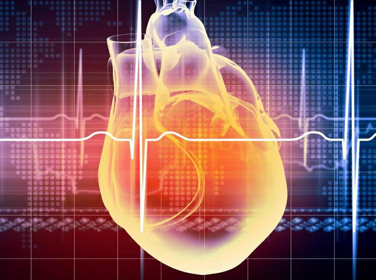
The atria are the two upper cavities inside the heart, the one on the left is called the "left atrium" and the one on the right is called the "right atrium", with thick walls and strong muscles. The atrium is a reservoir of blood and acts as an auxiliary pump. Ventricle is muscle pump, complete ejection function, muscle is strong.
The heart, like my fist and shaped like a peach, sits above my diaphragm and to the left between my lungs. The heart is a hollow muscular organ, mainly composed of myocardium, left atrium, left ventricle, right atrium, right ventricle four chambers. Between the left and right atria and between the left and right ventricles are separated by the septum, so they are not connected, there are valves between the atrium and the ventricle, these valves make the blood can only flow from the atrium into the ventricle, and can not flow back.
The one on the upper left is called the "left atrium" and the one on the upper right is called the "right atrium", which is thick and muscular. The left atrium is connected with the pulmonary vein, and the right atrium is connected with the venous phase of the upper and lower lumen and the opening of the coronary sinus. The left atrium receives blood from the lungs and the right atrium receives blood from the rest of the body.
There is a valve between the atrium and the ventricle, and when the atrium contracts, blood flows into the ventricle through this path. Blood flows from the atria into the ventricles, where it is pressed into the arteries and transported to the lungs and the rest of the body.
The heart, like my fist and shaped like a peach, sits above my diaphragm and to the left between my lungs. The heart is a hollow muscular organ, mainly composed of myocardium, left atrium, left ventricle, right atrium, right ventricle four chambers.
Between the left and right atria and between the left and right ventricles are separated by the septum, so they are not connected, there are valves between the atrium and the ventricle, these valves make the blood can only flow from the atrium into the ventricle, and can not flow back.
The one on the upper left is called the "left atrium" and the one on the upper right is called the "right atrium", which is thick and muscular. The left atrium is connected with the pulmonary vein, and the right atrium is connected with the venous phase of the upper and lower lumen and the opening of the coronary sinus. The left atrium receives blood from the lungs and the right atrium receives blood from the rest of the body.
There is a valve between the atrium and the ventricle, and when the atrium contracts, blood flows into the ventricle through this path. The part of the atrium is amazing, if sharp things stab into it, it will not be life-threatening, which is called not dead knot, but before the blood dries.
The blood is forced from the atrium into the ventricle, and from the ventricle into the artery, respectively, to the lungs and the rest of the body.
The right atrium is located in the upper right of the heart, the wall is thin and the cavity is large, and the part protruding from the left front is called the right auricle.
According to the direction of blood, the right atrium has three entrances and one exit: the upper orifice of the superior vena cava, the main body has the inferior vena cava, and the coronary sinus orifice between the inferior vena cava and the right atrioventricular orifice respectively guide the blood from the upper body, lower body and heart into the right atrium. The exit is the right atrioventricular orifice, through which the blood from the right atrium flows into the right ventricle.
In the lower part of the atrial septum of the right atrium there is a shallow oval fossa called the oval fossa. During fetal life, this is the foramen ovale, through which the left and right atria communicate. After birth, this hole gradually closed, and the remaining depression is called the oval fossa. If this hole is not closed about 1 year after birth, it will form a congenital disease - atrial septal defect (ovale atresia), accounting for 51% of congenital cardiovascular diseases.
The left atrium is located at the left rear of the right atrium and constitutes the majority of the bottom of the heart, and the part that protrudes to the right front is called the left auricle. The left atrium has four entrances and one exit: the entrance is the pulmonary vein, and the exit is the anterior and lower left ventricular opening, through which the blood from the left atrium flows to the left ventricle.
Atrial fibrillation (Af) is one of the most common arrhythmias, which is caused by many small reentry rings caused by the atria leading reentry ring.
(1) The symptoms of persistent (or chronic) atrial fibrillation are related to underlying heart disease, as well as to ventricular rate. There may be palpitations, shortness of breath, chest tightness, fatigue, especially after physical activity significantly increased ventricular rate, and may appear syncope, especially in elderly patients, due to cerebral hypoxia and vagal hyperactivity.
(2) Irregular heart rhythm: the first heart sound is uneven in intensity and interval. The ventricular rate of untreated atrial fibrillation generally ranges from 80 to 150 beats/min and rarely exceeds 170 beats/min.
Heart rate >100 beats/min, called rapid atrial fibrillation; >180 beats/min called extreme atrial fibrillation. Shortness of pulse.
(3) can induce heart failure or aggravate the original heart failure or basic heart disease, especially when the ventricular rate exceeds 150 times/time, can aggravate the symptoms of myocardial ischemia or induce angina pectoris.
(4) Increased susceptibility to thrombosis, and thus prone to embolism complications.
If atrial fibrillation lasts for more than 3 days, there will be thrombosis in the atrium. Older age, organic heart disease, enlarged left atrial diameter and increased plasma fibrin were risk factors for thromboembolic complications

